- 0086-571-85302990
- sales@greenskybio.com


The Role of 5-Aminolevulinic Acid in Brain Tumor Surgery: A Systematic Review
2025-09-27
Over the past few decades, advancements in medical technology have significantly transformed the landscape of brain tumor surgery. One of the promising innovations contributing to precision in neurosurgery is the use of 5-Aminolevulinic acid (5-ALA). Widely recognized for its role in enhancing tumor visualization, 5-ALA has become a pivotal tool in optimizing surgical outcomes by aiding the identification and resection of malignancies. This article offers a systematic review of how 5-ALA is utilized in brain tumor surgeries, examining its mechanisms, benefits, limitations, and impact on surgical practices.
Understanding 5-Aminolevulinic acid
5-Aminolevulinic acid (5-ALA) is a precursor in the heme biosynthesis pathway and is often employed as an intraoperative imaging agent. When administered orally prior to surgery, 5-ALA is preferentially taken up by malignant cells within the brain. It induces the accumulation of protoporphyrin IX (PpIX), a fluorescent compound visible under specific wavelengths of light. During surgery, surgeons use special blue-light filters in surgical microscopes, which cause the PpIX within tumor cells to emit a pink-red fluorescence. This fluorescence enhances the visualization of tumor margins, enabling surgeons to distinguish tumor tissue from normal brain tissue.
Mechanisms of Action
The mechanism by which 5-ALA enhances tumor visualization is rooted in its interaction with cellular metabolism. Tumor cells have alterations in their porphyrin metabolism, leading to greater accumulation of PpIX compared to normal cells. This differential accumulation results in fluorescence under blue light, aiding in the explicit delineation of tumor boundaries. The specificity of 5-ALA is particularly beneficial in glioblastoma multiforme (GBM), where tumor cells infiltrate surrounding brain tissue, making clear distinction crucial for effective resection.
Benefits of Using 5-ALA in Brain Tumor Surgery
Improved Tumor Resection: The primary advantage of 5-ALA is its ability to improve tumor resection rates. By allowing surgeons to see precise tumor margins, it facilitates more complete removal of tumor tissue while preserving healthy brain matter. Studies have reported increased rates of complete resection, which correlate with improved patient outcomes and survival rates.
Enhanced Visual Contrast: 5-ALA-induced fluorescence provides superior visual contrast during surgery, especially in complex brain tumors. This enhanced visualization is critical in distinguishing infiltrative tumor cells that otherwise blend into normal tissue during conventional white-light microscopy.
Reduced Risk of Recurrence: By achieving more thorough tumor removal, 5-ALA can decrease the likelihood of residual tumor tissue, potentially reducing recurrence rates. In high-grade gliomas, even small amounts of residual tumor can lead to rapid progression; thus, maximizing resection can be pivotal in delaying recurrence.
Improved Surgical Decision Making: The fluorescence guides surgical planning and execution, allowing surgeons to make informed decisions regarding the extent of resection while minimizing damage to vital neurological functions.
Increased Safety Profile: Despite its application, 5-ALA is generally well-tolerated among patients, with minimal adverse effects reported in clinical settings. This safety profile enhances its utility as a standard part of intraoperative procedures.
Limitations and Considerations
Despite its advantages, there are some limitations and considerations associated with the use of 5-ALA:
False Positives and Negatives: While 5-ALA provides enhanced visualization, there can be instances of false positives, where non-tumor tissues fluoresce, or false negatives, where tumor tissue does not produce detectable fluorescence. Such scenarios necessitate careful interpretation and reliance on a multidisciplinary surgical approach.
Efficacy Across Tumor Types: The effectiveness of 5-ALA varies among tumor types, with higher reliability observed in high-grade gliomas compared to low-grade variants. Its utility in non-glioma brain tumors may be limited, requiring customized strategies for visualization.
Costs and Accessibility: The integration of 5-ALA into surgical practice requires access to specialized equipment for fluorescence-guided resection, posing potential barriers in resource-limited settings. Additionally, the costs associated with the use of 5-ALA and necessary technology can be prohibitive.
Interdisciplinary Coordination: Successful deployment of 5-ALA requires coordination between surgical teams, anesthesiologists, and perioperative personnel, emphasizing the need for comprehensive training and collaboration.
Conclusion
5-aminolevulinic acid has emerged as a transformative tool in brain tumor surgeries, offering a detailed view of tumor margins and enabling more complete resection. Its application enhances surgical precision, contributing significantly to improved patient outcomes, especially in aggressive malignancies like glioblastomas. While challenges exist in terms of accuracy, access, and cost, ongoing research continues to unlock its potential as a pivotal component in neurosurgical practice. By forging pathways to overcome these limitations, 5-ALA is poised to remain at the forefront of advancements in neurosurgical oncology. Through systematic reviews and clinical trials, the medical community seeks to maximize the benefits of this innovative approach, ultimately shaping the future of brain tumor treatment for optimized patient care.


- ▶ Hesperidin
- ▶ Citrus Bioflavonoids
- ▶ Plant Extract
- ▶ lycopene
- ▶ Diosmin
- ▶ Grape seed extract
- ▶ Sea buckthorn Juice Powder
- ▶ Fruit Juice Powder
- ▶ Hops Extract
- ▶ Artichoke Extract
- ▶ Mushroom extract
- ▶ Astaxanthin
- ▶ Green Tea Extract
- ▶ Curcumin
- ▶ Horse Chestnut Extract
- ▶ Other Product
- ▶ Boswellia Serrata Extract
- ▶ Resveratrol
- ▶ Marigold Extract
- ▶ Grape Leaf Extract
- ▶ New Product
- ▶ Aminolevulinic acid
- ▶ Cranberry Extract
- ▶ Red Yeast Rice
- ▶ Red Wine Extract
-
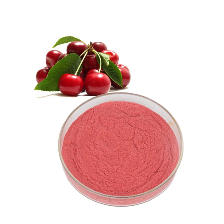
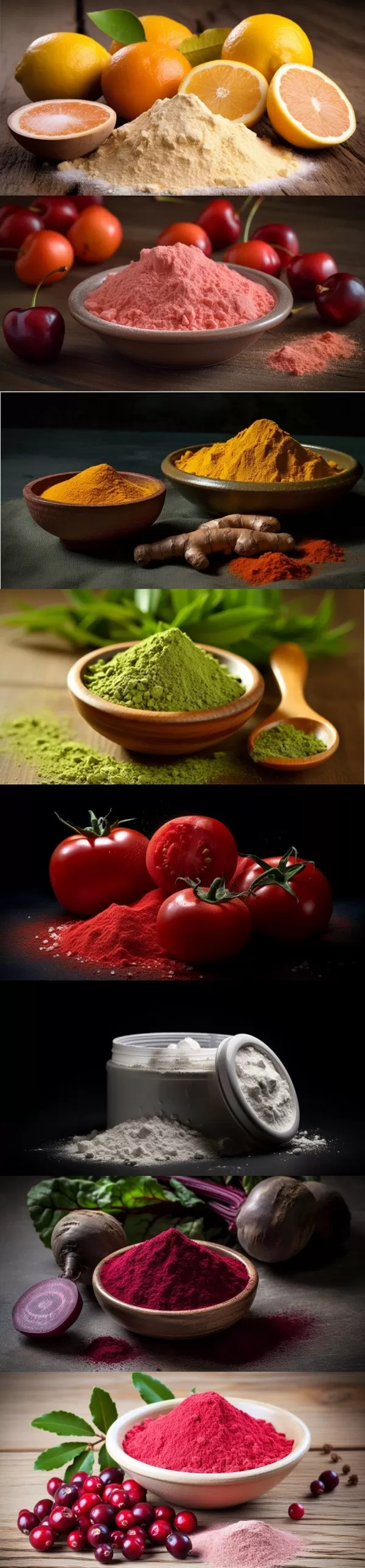
Acerola Extract
2025-09-27
-
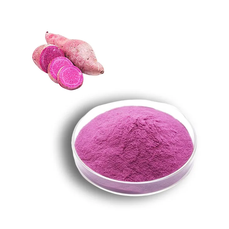
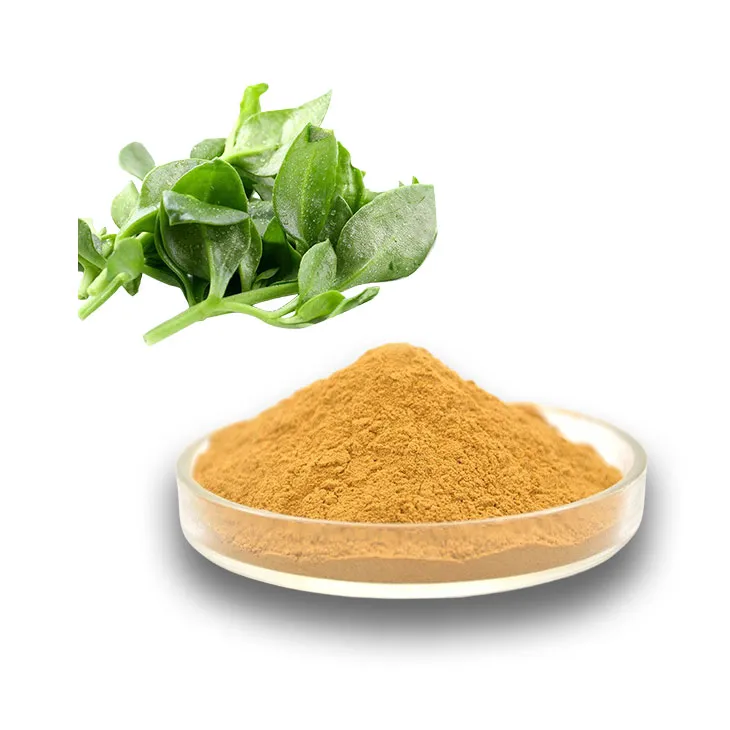
Purple Sweet Potato Extract
2025-09-27
-
Andrographis Paniculata Extract Powder
2025-09-27
-
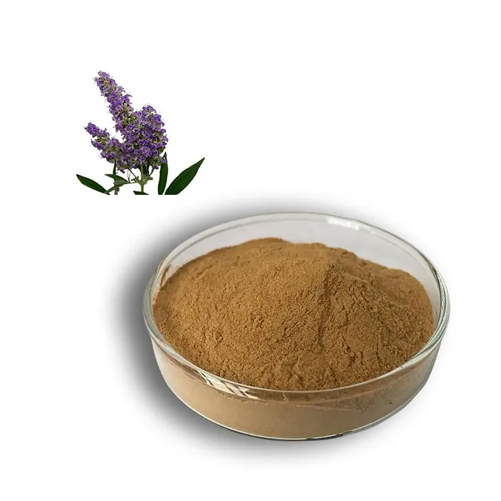
Chasteberry Extract
2025-09-27
-
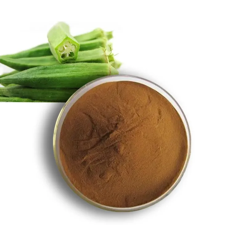
Okra Extract
2025-09-27
-
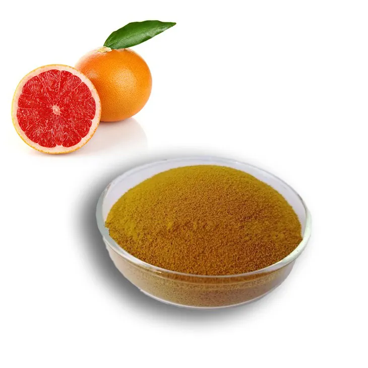
Grapefruit Seed Extract Powder
2025-09-27
-
Propolis Extract Powder
2025-09-27
-
Natural grape seed extract
2025-09-27
-

Red Wine Extract
2025-09-27
-
Hawthorn powder
2025-09-27
