- 0086-571-85302990
- sales@greenskybio.com


The Role of 5-Aminolevulinic Acid in Brain Tumor Surgery: A Systematic Review
2025-09-27
Aminolevulinic acid (ALA), a naturally occurring delta-amino acid, has become increasingly important across medicine, especially in cancer diagnostics and therapy, dermatology, and photodynamic treatments. Although its clinical utility is impressive, its effectiveness is rooted in a precise and elegant biochemical mechanism that capitalizes on both human and cellular metabolism. This article explores in detail the underlying mechanism of action of Aminolevulinic acid and its importance in diagnostic and therapeutic settings.
Introduction to Aminolevulinic acid (ALA)
Aminolevulinic acid, often referred to as 5-aminolevulinic acid (5-ALA), is a non-proteinogenic amino acid found in all nucleated cells. It is an essential precursor in the heme biosynthesis pathway—a process fundamental to cellular respiration, oxygen transport, and a host of biological functions. The critical importance of ALA does not stop at its role in physiology; synthetic and exogenous ALA forms the core of modern photodynamic therapy (PDT) and diagnostic fluorescence-guided surgery.
Endogenous Biochemical Pathway: Heme Biosynthesis
ALA as a Heme Precursor
ALA is the first committed precursor in the biosynthesis of heme, a component of hemoglobin, myoglobin, cytochromes, and various heme-containing enzymes. In human cells, ALA is synthesized from glycine and succinyl-CoA via the enzyme ALA synthase, largely in the mitochondria of liver cells and erythroblasts.
Simplified Steps:
Glycine + Succinyl-CoA → (via ALA synthase) → ALA
Two molecules of ALA condense (via ALA dehydratase) to form porphobilinogen.
Series of transformations lead to the formation of protoporphyrin IX (PpIX).
In the presence of ferrous iron (Fe2+), protoporphyrin IX is converted to heme by the enzyme ferrochelatase.
Regulation of Heme Biosynthesis
Heme production is tightly regulated through negative feedback to avoid toxic accumulation of pathway intermediates. Excessive accumulation—especially of PpIX—can lead to photosensitivity, which clinicians leverage in certain therapies.
Pharmacologic and Clinical Use of ALA
Exogenously administered (pharmaceutical) ALA is used for clinical purposes, most notably in:
Photodynamic therapy (PDT): For skin cancers, actinic keratosis, and certain neoplasms
Fluorescence-guided resection: Of high-grade gliomas and bladder tumors
Diagnostic cytology and histology
The unique capacity of exogenous ALA to accumulate in target tissues sets the stage for its diagnostic and therapeutic impact.
Mechanism of Action in Photodynamic Therapy (PDT) and Diagnosis
Topical or Systemic Administration
ALA is delivered either topically (for superficial lesions) or orally/intravenously (for systemic or deep tissue effects), where it penetrates target cells but is more avidly taken up by rapidly dividing or neoplastic cells compared to normal tissues.
Intracellular Conversion to Protoporphyrin IX (PpIX)
After cellular uptake, ALA enters the heme biosynthetic pathway but, in target (typically malignant or dysplastic) cells, the final conversion step (from PpIX to heme) is defective or downregulated. The rate-limiting enzyme, ferrochelatase, is often less active in cancer cells, resulting in selective accumulation of PpIX.
PpIX as a Photosensitizer
PpIX is an intensely fluorescent compound that absorbs light in the visible spectrum (especially at 405, 510, and 635 nm) and emits red fluorescence. Its ability to act as a photosensitizer is central to its clinical application.
In fluorescence-guided surgery, tumor tissues that have accumulated PpIX can be visualized using specific wavelengths of light, helping surgeons to distinguish tumor boundaries from normal tissue.
In PDT, after sufficient PpIX accumulation, the targeted tissue is illuminated with a light source matching PpIX’s absorption peak (commonly red light at 630-635 nm for deeper penetration).
Light Activation and Cytotoxic Cascade
Upon illumination, PpIX absorbs the light energy and undergoes a transition to an excited state. The excited PpIX then interacts with molecular oxygen in the tissue, generating reactive oxygen species (ROS)—primarily singlet oxygen (^1O_2).
ROS are highly cytotoxic and induce:
Direct damage to cell membranes (lipid peroxidation)
Protein denaturation and enzyme inactivation
DNA and RNA breaks or modifications
Disruption of cellular structures, vasculature, and mitochondrial membranes
These effects ultimately result in apoptosis (programmed cell death), necrosis, or autophagy of targeted cells.
Selective Damage and Therapeutic Specificity
Because ALA-induced PpIX selectively accumulates in abnormal or malignant tissue and the ROS have a limited sphere of influence (short diffusion distance), the cytotoxicity is spatially confined. This allows for destruction of diseased tissue with minimal damage to adjacent healthy cells.
Vascular and Immune Effects
Secondary effects of ALA-PDT include damage to tumor vasculature, leading to ischemia of lesions, and a local inflammatory response that may enhance anti-tumor immunity.
Applications in Clinical Practice
A. Dermatology
Actinic keratosis: Topical ALA-PDT is FDA-approved, providing high efficacy for pre-cancerous skin lesions with excellent cosmetic outcomes.
Superficial basal cell carcinoma and other skin neoplasms: ALA-PDT is an established therapy for selected cases, particularly where surgery is contraindicated.
B. Oncology and Surgery
Fluorescence-guided neurosurgery: Orally administered 5-ALA enables visualization of high-grade gliomas; surgeons can more accurately resect tumors, reducing recurrence.
Bladder cancer diagnosis and resection: Intravesical ALA results in selective fluorescence of malignant urothelium, guiding biopsy and excision.
C. Gynecology and Other Fields
ALA-PDT is under evaluation for cervical neoplasia and other dysplastic or malignant lesions.
Why Does PpIX Preferentially Accumulate in Tumors?
The explanation relates to:
Increased uptake and esterification of ALA by tumor cells
Reduced ferrochelatase activity in neoplastic cells
Higher metabolic demands and altered mitochondrial function in cancers
Perfusion and perfusion-related delivery of ALA
Potential Side Effects of ALA Therapy
Photosensitivity: Patients must avoid bright light or sunlight after ALA administration until PpIX levels drop to normal.
Local inflammation or pain: At site of therapy/illumination.
Rare systemic effects: Such as transient liver enzyme elevation.
Future Directions and Research
Research continues on:
Improving delivery systems for deeper tissues
Enhancing selectivity and cytotoxicity
Combining ALA-PDT with other chemotherapeutics or immunotherapies
Conclusion:
The mechanism of aminolevulinic acid centers on its biochemical role in the heme pathway, exploited by clinicians to induce the selective accumulation of protoporphyrin IX in target tissues. When activated by specific wavelengths of light, PpIX acts as a photosensitizer, generating cytotoxic ROS that destroy abnormal or malignant cells with notable precision. This dual role—selective diagnostic fluorescence and therapeutic cytotoxicity—makes ALA a unique and potent agent in modern photodynamic therapy and fluorescence-guided surgery. As research and technology advance, ALA is poised for even greater impact in the future of personalized medicine and minimally invasive interventions.


- ▶ Hesperidin
- ▶ Citrus Bioflavonoids
- ▶ Plant Extract
- ▶ lycopene
- ▶ Diosmin
- ▶ Grape seed extract
- ▶ Sea buckthorn Juice Powder
- ▶ Fruit Juice Powder
- ▶ Hops Extract
- ▶ Artichoke Extract
- ▶ Mushroom extract
- ▶ Astaxanthin
- ▶ Green Tea Extract
- ▶ Curcumin
- ▶ Horse Chestnut Extract
- ▶ Other Product
- ▶ Boswellia Serrata Extract
- ▶ Resveratrol
- ▶ Marigold Extract
- ▶ Grape Leaf Extract
- ▶ New Product
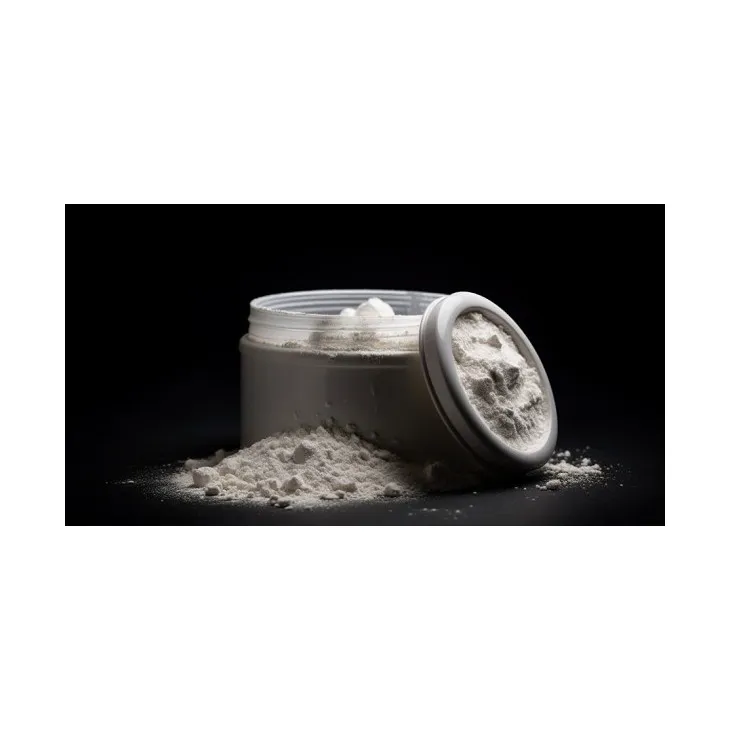
- ▶ Aminolevulinic acid
- ▶ Cranberry Extract
- ▶ Red Yeast Rice
- ▶ Red Wine Extract
-
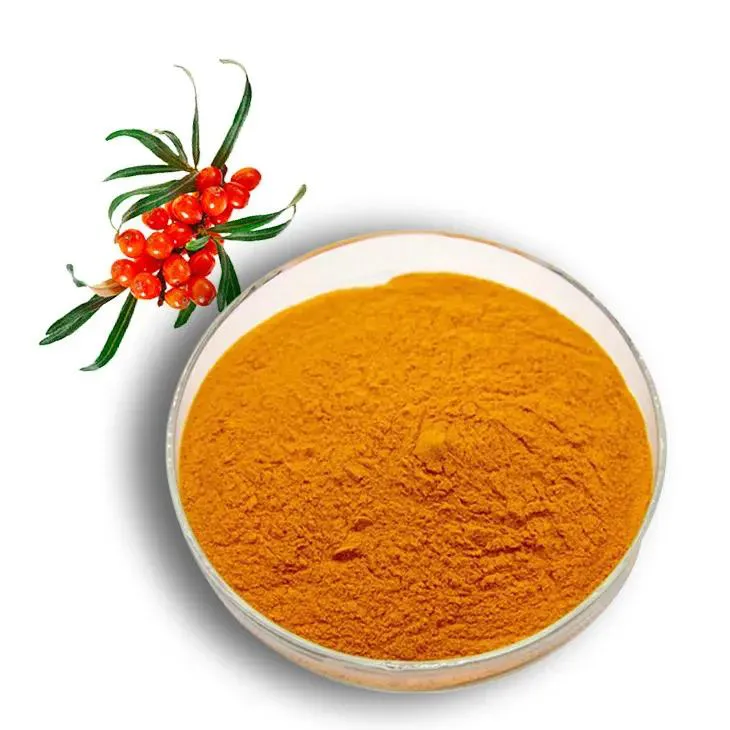
Sea buckthorn Juice Powder
2025-09-27
-
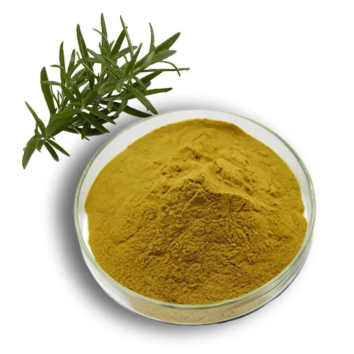
Rosemary extract
2025-09-27
-
Lavender Extract
2025-09-27
-
Golden Seal Extract
2025-09-27
-
Shikonin
2025-09-27
-
melatonin
2025-09-27
-
Clove Powder
2025-09-27
-
Sea buckthorn oil
2025-09-27
-
Black Garlic Extract
2025-09-27
-
Tamarind extract powder
2025-09-27
